Aortic Stenosis
What is it?
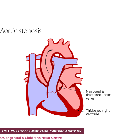
Aortic stenosis (or AS) is a condition where there is narrowing of the main artery to the body (the aorta) from the heart. The exact site of narrowing can be immediately below, at or above the aortic valve.
The normal aortic valve has three components, which are known as valve leaflets. Thickening of these leaflets or the presence of only two (known as bicuspid), or one, leaflet(s) can cause a narrowed valve.
The type of aortic stenosis that involves narrowing below the valve (known as subaortic stenosis) can be caused by increased muscle or it can be a discrete shelf of what is called 'fibrous' material.
All types of aortic stenosis will lead to a restriction of the outflow from the main pumping chamber of the heart, the left ventricle. This causes this muscle to become much thicker than normal (hypertrophy) as it works harder to pump the blood.
How many people get it?
The prevalence of aortic stenosis has been estimated at five cases out of every 10,000 live births accounting for about 5% of all congenital heart disease.
Who gets it?
Anyone can get this condition. Whatever the cause of aortic stenosis, it is present from birth and probably from very early on in the pregnancy (possibly the 7th week after conception).
What are the signs and symptoms?
The disease can come to medical attention at any age. If severe, the baby may present in the newborn period with severe heart failure. This appears as breathlessness and sweating, particularly whilst feeding. Less severe cases will be picked up with a heart murmur during the discharge baby check or 6 week check.
In older children, presentation may be due to the presence of a murmur on routine health screening. People tend not to have any symptoms, although they may complain of breathlessness, chest pain, fainting or dizziness on exercise. Unfortunately, some people who die suddenly are found to have severe aortic stenosis.
With the advent of ultrasound scanning of the unborn child's heart (fetal echocardiography), the diagnosis can sometimes be made before the child is born. This allows arrangements to be made for the infant to be seen in a cardiac centre after birth.
What kind of tests might I have?
Infants are usually referred to a paediatric cardiologist, who will investigate further. The diagnosis is made by echocardiography. This ultrasound scan will show that the valve is not opening properly. A chest X-ray and electrocardiogram (ECG) may be performed.
What is the treatment?
Mild and moderate aortic stenoses usually only need to be observed. If it is severe or is causing symptoms, then treatment is needed. If it is severe in a baby, attempts are made to improve the general state of the baby. There are two types of treatment for narrowing of the valve
- Balloon valvuloplasty at cardiac catheterisation. If treatment is needed and the narrowing is at the level of the valve then the valve can be directly ballooned internally. Essentially a catheter is inserted through the leg artery under general anaesthesia and a balloon placed across the valve. When inflated the valve stretches up. The catheter and balloon are then removed.
- Surgery
If the narrowing is above or below the valve, then the treatment is generally surgical.
What is the prognosis?
The prognosis is generally good and in the long term most children fare well leading normal lives without symptoms. Some patients require a second operation some time after the first. This can be a repeat balloon or surgical procedure. Sometimes the narrowed valve becomes leaky after an operation and this itself can be a problem. Where the valve is severely abnormal it may need to be replaced either by the pulmonary valve (Ross procedure) or by an artificial valve. Lifelong blood-thinning medicine is then required, along with regular monitoring blood tests.
Further information at The Children's Heart Federation