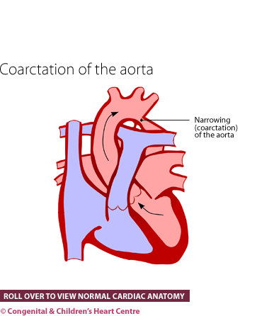
Coarctation of the aorta
What is it?
Coarctation of the aorta is a condition whereby there is an often severe narrowing in the main blood artery to the body (called the aorta). It occurs in the last part of the aortic arch.
How many people get it?
It has been estimated that there are five cases of coarctation out of every 10,000 live births, and this accounts for about 6% of all congenital heart disease.
What are the signs and symptoms?
The condition can be noticed in the newborn period or considerably later, in childhood or even, adulthood. In the newborn type of coarctation, the baby is usually well for the first day or so and then becomes suddenly (acutely) unwell. The baby will not feed, become breathless and a grey colour. Without help, the child will deteriorate rapidly. Sometimes, the condition is detected early on the routine discharge baby check, as the pulses in the legs may not be felt.
In the older child or adult, weakly felt pulses at a routine health check may be the sign that brings the condition to medical attention. In addition, it can be detected because of high blood pressure. These children and adults often appear to be very well.
With the advent of ultrasound scanning of the unborn child's heart (fetal echocardiography), the diagnosis can sometimes be made before the child is born. This allows arrangements to be made for the infant to be seen in a cardiac centre after birth, though it is usually not necessary for the child to be born in the cardiac centre itself.
What kind of tests might I have?
Infants and children are usually referred to a paediatric cardiologist and adults are referred to a congenital cardiologist, both of whom will investigate further. In the case of children, the doctor will question the parents to obtain the child's history and examine the child and then perform several tests.
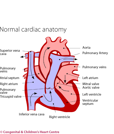
These will usually consist of an electrocardiogram (ECG) to measure the electrical activity of the heart and a chest X-ray to visualise the heart and lungs. The diagnosis is made by echocardiography. This ultrasound scan will show that there is a narrowing in the aorta, the main blood vessel to the body. Occasionally, further imaging is needed such as cardiac catheterisation and body scans such as magnetic resonance imaging (MRI) or computerised tomography (CT) that can give very good images of the aortic arch. Some blood tests may also be necessary.
What is the treatment?
- Drug therapy for newborns. In the case of sick newborn babies with this condition, the first treatment is to stabilise the child. This can involve using drugs to stimulate the heart and a medicine called prostaglandin. This restores the fetal circulation and stabilises the baby prior to surgery, which is the mainstay of treatment. The baby is then transferred to the cardiac unit.
- Surgery to remove the narrowing of the blood vessel.
- Balloon angioplasty at cardiac catherisation. This may be performed in older children and adults as a primary procedure. A stent may be inserted in the narrowed region to prevent the narrowing from recurring.
What is the prognosis?
The prognosis is usually extremely good. In the long term, these children fare well and most are have no symptoms and lead normal lives. Some require a repeat procedure which may be surgical or by balloon.
Download Coarctation of the Aorta PDF![]()
Further information at The Children's Heart Federation