Atrial Septal Defect and Patent Foramen Ovale

What is it?
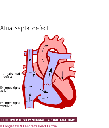
An atrial septal defect (or ASD) is a hole in the wall (septum) that divides the left and right collecting chambers of the heart (atria). This hole or defect in the septum allows the two collecting chambers (right and left atrium) to be in contact with each other. The lesion is usually isolated but can occur in conjunction with other more complicated types of heart disease.
There are four different types depending on exactly where the defect is in the atrial septum. The most common form is the secundum ASD. Here, the hole is in the centre of the atrial septum, a region that is open in the womb, allowing blood to pass across freely. It should close off at birth with a tissue flap; failure to do so leaves a hole.
Patent foramen ovale - where the hole is small and still partly covered by the tissue flap, it is known as a patent foramen ovale or PFO.
Sinus venosus ASD - where the defect is situated near one of the great veins (superior vena cava- SVC or inferior vena cava- IVC) draining back to the heart, it is termed a sinus venosus ASD.
Primum ASD - another type is known as the primum ASD. This is a hole in the lower part of the atrial septum (wall) and occurs in conjunction with an abnormality of the valve that divides the left collecting chamber (left atrium) from the pumping chamber (left ventricle).
How many people get it?
It has been estimated that there are eight cases of ASD for every 10,000 live births and this accounts for about 8% of all congenital heart disease.
Who gets it?
Anyone can get an ASD. Whatever its cause, it is present from birth and, in some subtypes, possibly from very early on in the pregnancy (possibly the 7th week after conception). The type with some flap valve tissue remaining (patent foramen ovale or PFO) is present in most newborn infants but tends to seal off spontaneously over the first few weeks and months. A PFO may stay open in some cases but if small (< 3mm) it is inconsequential and should be regarded as a normal finding.
What are the signs and symptoms?
With an ASD, there may be no or only mild symptoms. Frequent chest infections can occur and children may be thinner and not as tall as normal. There may be a reduction in the child's energy levels and exercise tolerance. A murmur (abnormal heart sound) is usually present and may be detected on routine health checks. Very occasionally, infants have increased breathlessness and difficulty feeding. Often the child is completely asymptomatic.
Often the patient first visits the doctor with problems in adulthood. Adults going to their doctor in their twenties and thirties with this condition who have not had any treatment may have problems with their heart rhythm, breathlessness, heart failure and high pressure in the lungs.
What kind of tests might I have?
Infants are usually referred to a paediatric cardiologist, who will investigate further. After questioning the parents to obtain the child's history and examining the child, several tests will be organised. These will usually comprise an electrocardiogram (ECG) to measure the electrical activity of the heart and a chest X-ray to visualise the heart and lungs.
The diagnosis is made by echocardiography. This ultrasound scan will show the hole in the heart. Sometimes, a special type of echocardiogram is necessary. This is called a trans-oesophageal echo or TOE (TEE in the US). In this test, the ultrasound probe is passed down the gullet (oesophagus) to look at the heart from within. This is usually performed under sedation
What is the treatment?
Often, small ASDs or the flap tissue type (patent foramen ovale) will close spontaneously. This can take several years to happen and it is safe for the cardiologist to watch them closely throughout this period. Larger defects require closure.
- Device closure at cardiac catheterisation. The child or adult is anaesthetised and a needle put in to the blood vessel at the top of the leg. A narrow tube (called a catheter) is passed through the vein up into the heart. The device is passed up the centre of the tube and deployed across the hole. This is performed under trans-oesophageal guidance. The device remains in the heart and becomes incorporated into it.
- Surgical repair
What is the prognosis?
The prognosis is excellent for the secundum type of ASD, whatever procedure is used to close the hole (surgery or cardiac catheter). In the long term, these children lead normal lives with no symptoms or complaints. In situations where the hole is discovered and treated in later adulthood, some of the problems (particularly those with heart rhythm) can persist, despite treatment. For those with an ostium primum type of hole there can be longer term problems with the left sided valve due to leakage. This sometimes requires further operations or even replacement.
Download Atrial Septal Defect and Patent Foramen Ovale PDF![]()
Further information at The Children's Heart Federation

